Abstract
Introduction
Maternal embryonic leucine zipper kinase (MELK) is a novel oncogene, which exerts pivotal roles in maintenance of cancer stem cells. OTSSP167, an orally bioavailable inhibitor targeting MELK, is currently in a phase I/II clinical trial in patients with acute myeloid leukemia and breast cancer. Yet, no literature has been reported regarding the effects of MELK and OTSSP167 in chronic lymphocytic leukemia (CLL). Intriguingly, previous studies identified that OTSSP167 contributed to tumorigenesis via p53 pathway, highlighting its potential efficacy in relapsed/ refractory CLL. Hence, the aim of this study was to determine the anti-tumor potency and mechanism of OTSSP167 targeting MELK in CLL.
Methods
Peripheral blood samples from 55 de novo CLL patients were collected with informed consents at the Department of Hematology in Shandong Provincial Hospital Affiliated to Shandong University (SPHASU). CD19+ B cells were isolated with informed consents from healthy donors. Expression levels of MELK mRNA and protein in CLL cells were determined by quantitative RT-PCR and western blotting. Kaplan-Meier survival curves with log-rank test of overall survival (OS) were analyzed. Microarray datasets GES22762 and GSE25571 were obtained from Gene Expression Omnibus. Functional enrichment analyses of gene ontology (GO), kyoto encyclopedia of genes and genomes (KEGG) and gene set enrichment analysis (GSEA) in gene expression profiles were performed. Lentiviral vectors transfected CLL cells to stably silence MELK and adenovirus delivery were constructed to overexpress MELK. Besides, cell viability, apoptosis and cell cycle were assessed by cell counting kit-8, annexin V-PE/7AAD and PI/RNase staining, respectively. Cell migration was evaluated by CXCL12-induced chemotaxis.
Results
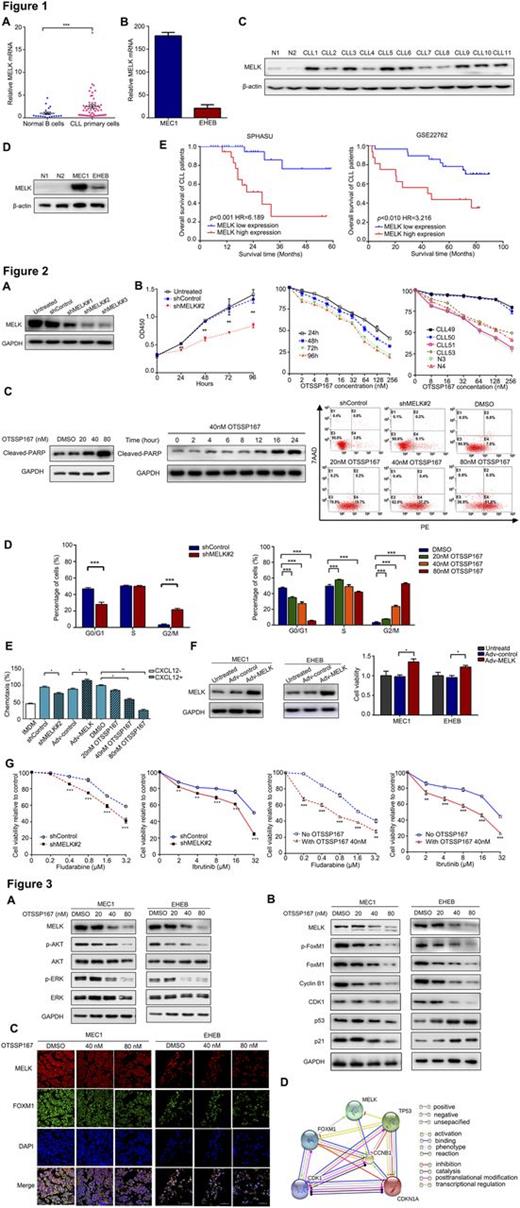
Aberrantly increased expression of MELK was detected in CLL primary cells and cell lines (MEC1 and EHEB) at mRNA and protein level compared with normal B cells (Figure 1A-D). We observed MELK over-expression in significant correlation with higher WBC count( p=0.022), advanced Binet stage (p=0.023), high-risk Rai stage (p=0.022), elevated LDH level (p=0.002) and increased β2-MG levels (p=0.009), unmutated IGHV (p=0.037), positive ZAP-70 (p=0.037) and deletion of 17p13 (p=0.009). Besides, MELK high expression was revealed in significant association with reduced OS of enrolled patients, which was further confirmed in GSE22762 (Figure 1E).
Annotations of bioinformatics analyses indicated that MELK was functional enriched in cell proliferation, regulation of G2/M cell cycle, response to drug and p53 signaling pathway in cancer. In consistent with functional enrichment analyses, CLL cells with silence or inhibition of MELK exhibited attenuated cell proliferation, increased fast-onset apoptosis, induced G2/M phase arrest, impaired cell chemotaxis and enhanced sensitivity to fludarabine and ibrutinib (Figure 2A-E). Whereas, gain-of-function assay showed promoted cell proliferation and cell chemotaxis (Figure 2E-F). Additionally, serial dilution of OTSSP167 decreased phosphorylation of AKT and ERK1/2 in CLL cells (Figure 3A). Besides, expression of phosphorylated FoxM1, FoxM1, cyclinB1 and CDK1 decreased with treatment of OTSSP167 in CLL cells. p53 modestly changed with MELK suppression in MEC1 cells, yet visibly elevated in EHEB cells. Besides, activation of p21 was observed (Figure 3B). Furthermore, confocal immunofluorescent images revealed that MELK and FoxM1 co-localized in CLL cells and were down-regulated by OTSSP167 in a dose-dependent manner (Figure 3C). Collectively, the interactive network of MELK and FoxM1-signaling cascade in genomic expression profile was established, illuminating the potential mechanism of MELK inhibition by OTSSP167 in CLL (Figure 3D).
Conclusion
Our investigations identified for the first time the oncogenic role of MELK in CLL tumorigenesis and regulatory mechanism of OTSSP167 in CLL cells by bioinformatics analysis and ex vivo evaluation. Expression of MELK was upregulated, and associated with adverse outcome of CLL patients. OTSSP167 exerted therapeutic potential at nanomolar concentrations in abrogating cell survival and migration, inducing cell cycle arrest through down-regulation of FoxM1. Further clinical investigation is warranted to substantiate OTSSP167 as a novel therapeutic strategy in progressed CLL.
No relevant conflicts of interest to declare.
Author notes
Asterisk with author names denotes non-ASH members.


This feature is available to Subscribers Only
Sign In or Create an Account Close Modal